Infection
Infectious diseases are caused by bacteria, fungi, viruses, and multicellular parasites (e.g., worms). The health significance of each of these agents is affected by many variables, some of which were important factors during the history of human disease.
For example, parasites may have become a greater problem for human groups after the domestication of animals, which are common vectors of diseases associated with these organisms. Airborne viruses, which require fairly large host populations (Fiennes 1978), may have become a significant health problem with the rise of urbanism. Fungi may have become a more serious problem with the advent of agriculture, particularly among groups who were under- or malnourished. Because fungi tend to be opportunistic, they are a greater problem in individuals who are immunologically compromised.Perhaps the most interesting observation about infectious disease during the Mesolithic through the Metal Ages is its apparent low incidence in skeletal samples from most geographic areas (e.g., Wood- Jones 1910b; Martin et al. 1984; Rathbun 1984; Strouhal 1986). This low frequency has been observed both for localized infection resulting from trauma in general (Domurad 1986) and fracture in particular (Wood-Jones 1910b; Bennike 1985) and for skeletal lesions (Martin et al. 1984), which may develop as a result of systemic disease. In fact, skeletal evidence of systemic infection appears to be quite rare, and differential diagnosis often poses a great challenge even to experienced paleopathologists or orthopedic specialists.
Even though skeletal studies report a low incidence of infection, interpreting this observation requires caution and reflects some of the theoretical problems involved in paleopathology. Most serious, acute infectious diseases leave no evidence in gross skeletal remains. The lack of skeletal evidence of infection may mean that the population was healthy, but it may also mean that people were dying from acute infectious disease before the skeleton could be affected (Ortner and Putschar 1981; Rathbun 1984; Ortner 1991).
Several surveys have detected possible trends in infection rates. Rathbun (1984) notes that in Iraq and Iran there appears to have been an increased frequency of infectious-type lesions in the Neolithic and Chalcolithic periods. These rates declined significantly in the later Bronze and Iron ages. Although these trends may be real, they may also reflect stochastic effects of inadequate samples. As Rathbun (1984) suggests, however, the decline in percentage of cases exhibiting infectious lesions from the Chalcolithic to the later Bronze and Iron Age periods may be the result of people dying before bone was involved. Elsewhere in a study of European skeletal material, Meiklejohn and colleagues (1984) noted an increase in cranial infections from the Mesolithic to the Neolithic period. Dental evidence of disease in the eastern Mediterranean suggests to Angel (1984) that new diseases, including epidemics, emerged with the increased population sizes of the Middle Bronze Age.
Evidence from surveys of archeological skeletal material, site reports, and individual case reports has pointed to the existence of a number of infectious diseases in the pre-Roman era - specifically, tuberculosis, leprosy (in Egypt), osteomyelitis, and periostitis.
There is strong evidence that tuberculosis may have developed and/or become a problem with the beginning of farming. Although no unequivocal cases of destructive lesions of the spine have been reported thus far for the prefarming Mesolithic, there are numerous descriptions of lesions of the spine for Neolithic Europe (c. 4200-1500 B.C.; Bartels 1907; Patte 1971; Sager et al. 1972; Dastugue and de Lumley 1976; Formicola, Milanesi, and Scarsini 1987) and pre-Dynastic Egypt (c. 48003100 B.C.; Strouhal 1991), for which one possible diagnosis is tuberculosis. However, one must caution that several disease conditions other than tuberculosis produce destructive lesions of the spine, including infection by staphylococcus bacteria and fungi.
There were several cultural changes during the Neolithic that would have influenced the development of diseases affecting the spine. In the Near East the use of domestic animals became an important part of the economy. Proximity to domestic animals, particularly bovids, was likely to have been a significant factor in the development of tuberculosis (Manchester 1983). In addition, a more sedentary life-style and increased population density in the Neolithic and subsequent periods would have been likely to increase its incidence. However, farming was also likely to have increased the incidence of fungal diseases. It is thus probable that spinal lesions attributable to both bacterial and fungal infection increased in the Neolithic period. The current skeletal evidence supports this expectation, although the specific etiology for the destructive lesions remains problematic.
Reported cases of tuberculosis show wide geographic distribution. K. Manchester (1984), basing his opinion on monographic as well as on skeletal evidence, believes that the earliest undisputed evidence for human tuberculosis comes from Egypt and that the disease spread out from a near eastern center (Manchester 1983, 1984). Although there is considerable controversy regarding the exact dating
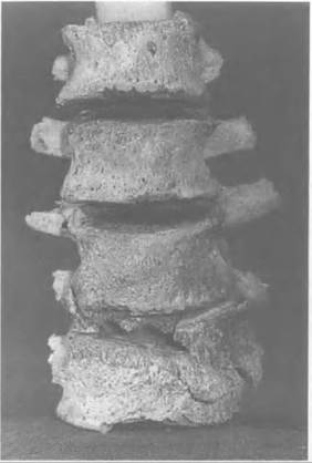
Figure V.1.2. Partial antemortem destruction of the fourth lumbar vertebral bone from the skeleton of a young adult from the Early Bronze Age cemetery (c. 3100 B.C.) at Bab edh-Dhra,, Jordan (Tomb A100E). Note the reactive bone formation on adjacent vertebral bodies, particularly the fifth lumbar vertebra. (Unaccessioned specimen, National Museum of Natural History, Washington, D.C.)
and diagnosis of the purported cases of spinal tuberculosis in Egypt (Morse 1967; Grmek 1989; Strouhal 1991), it appears that skeletal samples from this region contain at least 32 possible cases in the preChristian era (Strouhal 1991).
Donald Ortner (1979) reports two possible cases of tuberculosis in Jordan (Figure V. 1.2) in a sample of about 300 from the Early Bronze IA (c. 3100 B.C.) tombs at Bab edh- Dhra,. In central and southern Europe, in addition to the Neolithic cases already cited, five cases have been reported between the Bronze Age and 100 B.C. (Dastugue and de Lumley 1976; Steinbock 1976; Angel 1984). Manchester (1984) believes that in Britain the disease was present by the Roman period. Evidence of tuberculosis is also reported for northern Europe, where it may have begun later than the rest of Europe; most skeletal evidence is post-Roman (Brothwell, as cited in Manchester 1984).Leprosy is another infectious disease for which there is skeletal evidence. In a review of the history of leprosy, Manchester (1984) cites the earliest evidence as that from Ptolemaic Egypt (second century B.C.). Skeletal evidence for leprosy occurs later in Europe. D. Brothwell (1961) argues that leprosy was probably not established there before the first or second century A.D. and did not become a serious problem until after the seventh century. By the Mid
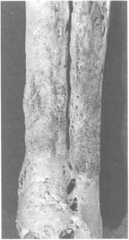
Figure V.1.3. Chronic inflammation of the left tibia and fibula resulting from an overlying ulcer in a skeleton of an adult woman probably over 50 years of age at the time of death. Specimen is from Tomb AlOOE in the Early Bronze Age cemetery at Bab edh-Dhra,, Jordan. Note the large, oval, circumscribed area of reactive bone associated with the ulcer. Chronic inflammation involves all bone tissue and resulted in fusion of the tibia and fibula. (Unaccessioned specimen, National Museum of Natural History, Washington, D.C.)
die Ages, the disease had become a serious problem but virtually disappeared by the end of the fifteenth century. Manchester (1991) notes that the reduced frequency of leprosy is associated with an increase in the prevalence of tuberculosis.
He speculates that a factor in the decline of leprosy may have been crossimmunity induced by the closely related disease of tuberculosis.Both osteomyelitis and periostitis have been reported in ancient skeletal material. Although their frequencies show some degree of variability (e.g., Wells, as cited in Sandison 1980; Strouhal 1986), these conditions generally are not common. Descriptions of cases are encountered in the literature. For example, Wood-Jones (1910b) describes a humerus from ancient Nubia with evidence of inflammatory bone formation. He attributes the inflammation to an infectious complication of an injury. Ortner (1979) reports a case of reactive bone formation on the tibia and fibula of a skeleton (Figure V.1.3) dated to about 3100 B.C. The specimen is of an old woman with an overlying skin ulcer of the lower leg that stimulated the inflammatory bone reaction.
Research on mummy remains has provided useful information on the antiquity of many other infectious diseases. An early case of poliomyelitis was reported in a pre-Dynastic mummy from Egypt (Mitchell 1900) dated to 3700 B.C. The leg deformity in the mummy of Pharaoh Siptah from the Nineteenth Dynasty (c. 1200 B.C.) has also recently been recognized as having been caused by polio (Fleming et al. 1980; Whitehouse 1980). Further possible evidence of this disease has been reported in Middle Bronze Age Greece (Angel 197 lb) and sixth- to second-century B.C. Italy (Mallegni, Fornaciari, and Tarabella 1979).
Mummy material has also provided information on parasites (Tapp 1986). Schistosomiasis is documented in two mummies of the Twentieth Dynasty (c. 1100 B.C.) in Egypt (Sandison 1973) but probably became a problem much earlier. The practice of irrigation, which may have begun in the Near East as early as the Neolithic, would have facilitated the spread of this disease. The roundworm, Ascaris, was found in an Egyptian mummy dating to around 170 B.C. (Fleming et al. 1980), as well as in coprolite material from Bronze Age Britain (Jones and Nicholson 1988) and Iron Age Europe (800-350 B.C.; Aspock, Flamm, and Picher 1973). The liver fluke was found in two mummies of the Twelfth Dynasty (Fleming et al. 1980). The tapeworm Echinococcus granulosus is associated with the mummy Asru dated to about 700 B.C. (Tapp and Wildsmith 1986).