79 Leishmaniasis
Leishmaniasis is primarily a skin disease produced by a number of different species of protozoa (genus Leishmania). The disease occurs in three basic clinical forms, each of which has several variants caused by different species, subspecies, or strains of the pathogen.
The intermediate host is the sandfly.Distribution and Incidence
Cutaneous leishmaniasis (often called “oriental sore”) is found in Armenia, Azerbaijan, Turkmenistan, Uzbekistan (republics of the former Soviet Union), Afghanistan, India, Iran, much of the Middle East and North Africa, the Sahara, the savanna states from Sudan to Senegal, and in Kenya and Ethiopia. In the New World, species of Leishmania cause various clinical forms of the disease in Central America, the Amazon Basin, the Guyanas, and the Andes, especially Venezuela and Peru. In eastern South America a form of the disease mainly afflicting children extends from Argentina to Venezuela and north through Central America to Mexico.
Mucocutaneous leishmaniasis is restricted to the New World and occurs in Brazil, eastern Peru, Paraguay, Ecuador, Colombia, and Venezuela.
Visceral leishmaniasis is found in India, Burma, Bangladesh, China, Thailand, Somalia, Chad, Kenya, Gabon, Sudan, and Niger. A variant occurring primarily among children is spread over southern Europe, North Africa, and the Middle East as well as Romania and the southern part of the former Soviet Union.
As a result of high levels of disease in rodent and dog populations, leishmaniasis is so common in endemic areas that it leaves its mark on every inhabitant. Recent estimates indicate that some 12 million individuals have one form or another of this infection. Thus leishmaniasis can be regarded as second in importance only to malaria among the protozoal diseases of humans. Although the mortality is low for the skin disease, it is almost always fatal for kala-azar, an organ variant.
Epidemiology and Etiology
All forms of leishmaniasis are zoonoses transmitted to human beings from wild or domestic animals via the sandfly (usually Phlebotomus). The leishmanial form of the parasite lives in reticuloendothelial cells of the mammalian host where it divides by binary fission to destroy the host cell. The parasites are taken up by the sandfly while feeding on the skin of the host, and in the insect’s intestine they develop into Ieptomonad forms. These divide, producing enormous numbers, and work their way to the pharynx and buccal cavity. There is a natural restriction of individual Ieishmaniae to specific sandflies, even though a wide variety of these insect species may be available.
Cutaneous leishmaniasis is caused by several members of the Leishmania tropica species complex. All produce chronic skin lesions, which tend to ulcerate. Some forms tend to be “urban” and closely linked to dogs as alternating hosts. Others are “rural,” with nonhuman reservoirs including various rodents, marsupials, and foxes. In the Americas, sandflies of the genus Lutzomyia are often vectors. The initial lesion usually heals spontaneously, but often leaves a disfiguring scar.
Mucocutaneous leishmaniasis is caused by Leish- mania braziliensis. In the form known as espundia in Brazil, the initial lesion develops into an infection of the mucosal tissues of the nose and mouth, resulting in gross deformities and sometimes death from secondary infections. Lutzomyia flies are the major vectors. (A clinically similar form, believed to be caused by L. tropica, has been described in Ethiopia.)
Visceral leishmaniasis, or kala-azar, is caused by at least three members of the Leishmania donoυani complex. In visceral leishmaniasis, unlike other forms of the disease, the organisms parasitize reticuloendothelial cells beyond the subcutaneous and mucosal tissue, and a number of internal organs may be involved. Symptoms include swelling of the liver and spleen, fever, diarrhea, emaciation, anemia, darkening of the skin, and gross abdominal enlargement.
Mortality in untreated cases has reached 75 to 95 percent over a 2-year period.Clinical Manifestations and Pathololgy
Old World cutaneous leishmaniasis has an incubation period of 6 weeks to a year, depending on the species of Leishmania. The lesions may be multiple, and it is thought that they are the result of multiple bites. There is much local induration and possible lymphatic spread to the regional lymph nodes. Healing is slow, with the formation of scar tissue in a year or so.
The New World disease takes several different forms:
1. A self-limiting ulcer of 6 months’ duration without metastases, caused by Leishmania tropica mexicana, is found in the Yucatan. It has a rodent reservoir in flat, low, rain forests.
2. Espundia, due to L. braziliensis, exists in the jungles ofPanama through Amazonia to Paraguay. It starts as a painless ulcer and metastasizes to the oronasal or anorectal area in 5 to 23 years. The host is thought to be the paca {Cuniculus paca), a nocturnal American rodent.
3. A solitary ulcerative lesion known as uta, usually found at the base of the nose with no metastasis, exists in the Andean valleys of Peru and Argentina. The agent is Leishmania peruviana, and the host is the dog.
4. Leproid leishmaniasis is seen in Venezuela. It begins as a fleshy cutaneous nodule that slowly spreads over the body without ulcerating and is difficult to distinguish from leprosy. It is thought to be due to an immune response and is very chronic.
In the absence of demonstrable Ieishmania, reliance rests on the Ieishmanin skin test, which is positive in cutaneous disease when there is a tuberculoid histology. Generally it becomes positive early and remains so after healing is complete. Although most lesions are self-limiting, antimony, pyrimethamine, and amphotericin B are used to treat metastases. A persistent positive serology is an important sign of continued survival of the parasite, which may live for decades in asymptomatic individuals.
History and Geography
Leishmaniasis is an ancient disease of both the Old and the New Worlds. Old World cutaneous leishmaniasis was first described in English by Alexander Russell in the mid-eighteenth century. Designs on Indian pottery of the New World clearly show the disfiguring disease. The pre-Columbian Incas knew of the dangers of the disease in the lowlands east of the Andes where coca grew and thus used captives to cultivate it. The Spaniards who later took over the coca trade were less aware of the problem, and consequently, their labor policies resulted in much disfigurement and death.
Visceral leishmaniasis was known to nineteenthcentury British doctors in India as kala-azar or Dumdum fever with its symptoms attributed vari-
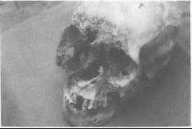
Figure VΠI.79.1. The uta form Ofleishmaniasis.
ously to malaria, Malta fever, and other diseases. It was in 1900 that W. B. Leishman, working at Net- tley, noticed the similarity of the parasite to that of trypanosomiasis, and shortly thereafter Leishman’s bodies were discovered to be the cause of kala-azar. Leishman published his findings in 1903, yet his work was duplicated independently by Charles Donovan. Leishman’s name was given to the entire genus, but the agent of kala-azar got its specific name from Donovan.
Old World leishmaniasis or oriental sore had long been in northern Africa and India, where it was known geographically as the Delhi boil, Aleppo boil, and so forth. Its agent, L. tropica, was probably seen as early as 1885, but the first clear description was not published until 1898, by Peter Fokitsch Borovsky in a Russian military journal. However, the paper did not become known to the West, and thus credit for the discovery of the organism is often given to James Homer Wright of Baltimore, who in 1903 found it in an ulcer of a child from Armenia.
The first cases of cutaneous disease in the Americas were described by A.
Carini and V Paranhos in southern Brazil in 1909 - the same year that mucocutaneous leishmaniasis was described as a distinct disease, this also in Brazil. Gasper Oliveira de Vianna named the etiologic agent Leishmania braziliensis in 1911. The visceral form in the Americas was first seen in Paraguay in 1913. Phlebotomus was suspected of being the vector as early as 1911, but this was not proven until 1941.The cutaneous and mucocutaneous form of the disease are relatively common problems among people working in heavily forested areas, such as the original area of Amazonia in Brazil. In Peru the uta form is seen on the lower eastern slopes of the Andes. In the Arica area of northern Chile there is an interesting case of what is probably the uta form (Figure VΠI.79.1) of this disease dating back about 1,000 years. It is known that contacts were common with the jungle area for trade purposes, and this case is probably an example of an imported exotic disease to the Pacific coast.
Marvin J. Allison
Bibliography
Gade, Daniel W. 1979. Inca and colonial settlement, coca cultivation and endemic disease in the tropical forest. Journal OfHistorical Geography 5: 263-79.
Herrer, Aristides, and Howard A. Christensen. 1975. Implication of Phlebotomus sand flies as vectors of bartonellosis and leishmaniasis as early as 1764. Science 190: 154-5.
Jones, T. C., et al. 1987. Epidemiology of America: Cutaneous leishmaniasis due to Leishmania braziliensis. Journal OfInfectious Diseases 156: 73-83.
Kean, B. H., Kenneth E. Mott, and Adair J. Russell, eds. 1978. Tropical medicine and parasitology: Classic investigations, Vol. I, 228-70. Ithaca.
Lainson, R., and J. J. Shaw. 1978. Epidemiology and ecology OfLeishmaniasis in Latin America. Nature {Parasitology Supplement) 273: 595—600.
Markell, Edward K., Marietta Voge, and David T. John. 1986. Medical parasitology, 6th edition. Philadelphia.
Strong, R. P. 1944. Stitts diagnosis, prevention and treatment of tropical disease. 7th edition. Philadelphia.
Vianna, Gaspar Oliveira de. 1911. Sobre o tratemento de Ieishmaniose tegumentar. Anais Paulistas de Medicina e Cirurgia 2: 167—9.